A Comprehensive Look at Skin Cancer Screening
Skin cancer, the abnormal growth of skin cells, is the most common form of cancer in the United States. Early detection is crucial for successful treatment. Skin cancer screening plays a vital role in identifying suspicious lesions early, improving the chances of a positive outcome. Here, we delve extensively into the world of skin cancer screening, empowering you to understand the process, its importance, and how to participate in protecting your skin health.
The Importance of Early Detection:
Skin cancer is highly treatable when detected and addressed promptly. Early-stage skin cancers are often curable with minimally invasive procedures. However, delayed diagnosis can lead to the need for more extensive surgery, potentially with significant scarring and functional limitations. In advanced stages, skin cancer can spread to other parts of the body, significantly impacting overall health and survival rates.
Who Should Get Screened?
The need for regular skin cancer screening depends on several factors:
- Age: The risk of skin cancer increases with age. Individuals over 50 are at a higher risk and should prioritize regular screenings.
- Skin Type: People with fair skin, those who burn easily and tan minimally, have a higher risk due to lower melanin production (melanin protects the skin from UV radiation).
- Sun Exposure: Excessive sun exposure throughout life is a major risk factor. People with a history of frequent sunburns or intense sun exposure are at increased risk.
- Personal History: A history of skin cancer or having numerous atypical moles (moles with unusual features) increases the need for regular screening.
- Family History: Having a close relative with skin cancer raises your risk.
Types of Skin Cancer Screening:
There are two primary approaches to skin cancer screening:
- Self-Examination: Regularly examining your skin at home is crucial for identifying any changes in existing moles or the appearance of new lesions.
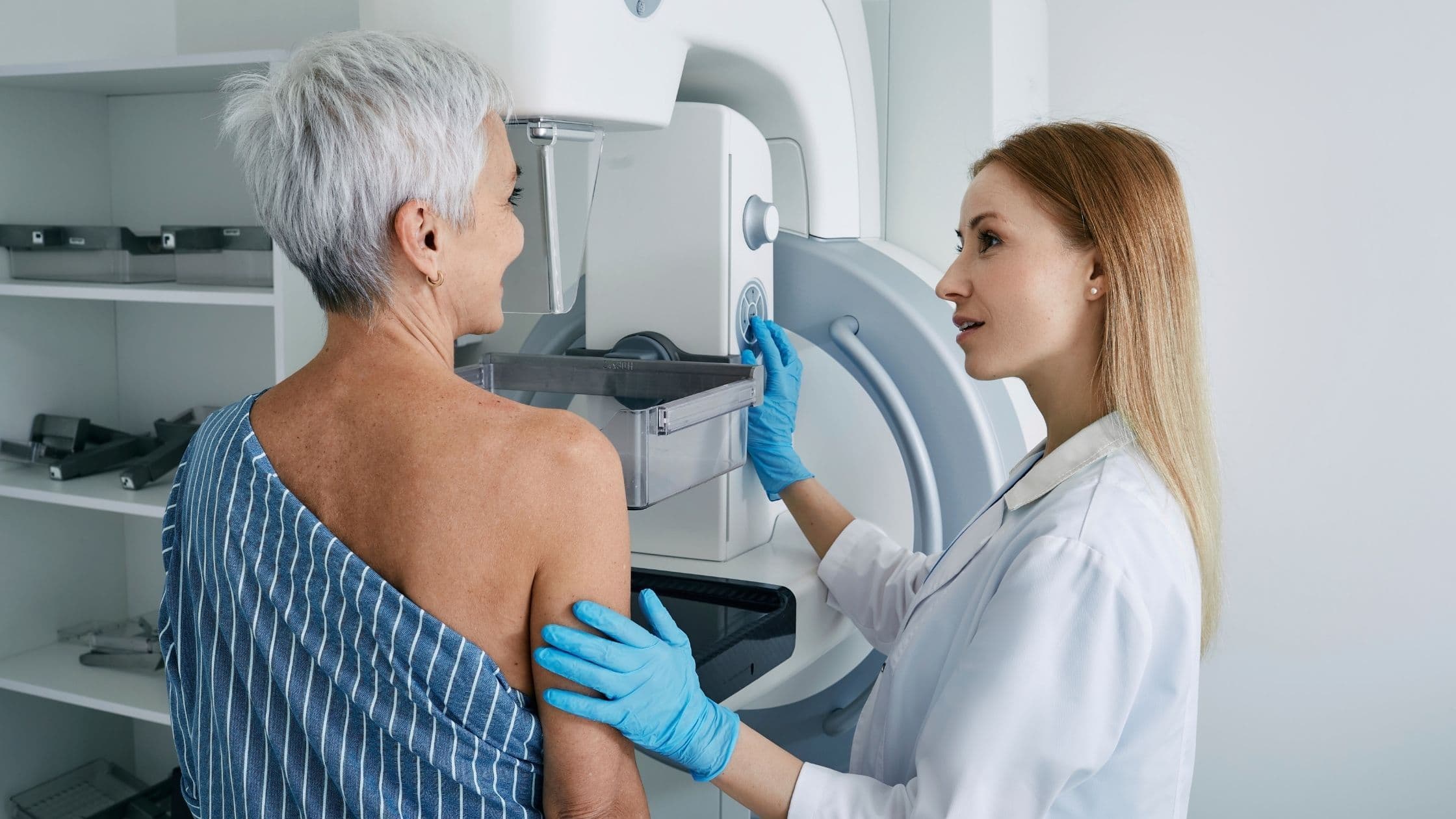
- Clinical Examination: A healthcare professional (HCP), typically a dermatologist, performs a comprehensive skin exam to check for suspicious lesions.
Self-Examination:
Here's a guide for performing a thorough self-examination at home, ideally once a month:
- Utilize a Mirror: Examine your entire body using a full-length mirror and a handheld mirror for areas that might be difficult to see on your own.
- Focus on All Areas: Pay close attention to areas exposed to the sun, including your face, ears, scalp (parting your hair), neck, chest, arms, legs, and back. Don't forget to check your palms, soles of your feet, and genitals.
- Look for Changes: Be aware of any changes in the size, shape, or color of existing moles. Also, watch for new moles or any unusual spots, bumps, or patches of discolored skin.
- Use the ABCDE Rule: This can help you memorize the warning signs of suspicious moles:
- Asymmetry: One half of the mole doesn't match the other half.
- Border: The borders are irregular, ragged, or blurred.
- Color: The mole has multiple colors within it, such as brown, black, red, white, or blue.
- Diameter: The mole is larger than 6 millimeters (about the size of a pencil eraser).
- Evolving: The mole is changing in size, shape, or color.
Clinical Examination by an HCP:
During a clinical examination, your HCP will:
- Ask about your medical history and risk factors.
- Perform a full-body skin exam, meticulously examining your skin from head to toe. They might use a dermatoscope, a magnifying instrument with a light source, to get a closer look at suspicious lesions.
- Discuss any concerns and recommend further evaluation if necessary. This might involve dermoscopy imaging or biopsy, where a small sample of tissue is extracted for microscopic examination to confirm or rule out skin cancer.
Additional Screening Techniques:
In some cases, depending on your individual risk factors, your HCP might recommend additional screening methods:
- Dermoscopy Imaging: This non-invasive technique utilizes a dermatoscope to capture high-resolution digital images of suspicious lesions. These images can be compared over time to monitor for changes or used for specialist consultation.
- Mole Mapping: This involves creating a detailed photographic map of your body's moles, documenting their location, size, and appearance. This can be helpful for tracking changes over time.
The Importance of Follow-Up:
Following up with your HCP after a screening is crucial. They will discuss the results and recommend appropriate next steps. This might involve monitoring suspicious lesions, scheduling follow-up appointments, or referring you to a dermatologist